이분 척추(척추 피열),잠제성 이분 척추(폐쇄성 이분 척추),수막류(수막 허니아/뇌수막 탈출증)와 척수 수막류, Spina bifida, Spina bifida occulta, Meningocele and Myelomeningocele
이분 척추의 종류
- 이분 척추에는
-
- 잠재성 이분 척추,
- 수막류,
- 척수 수막류가 있다.
- 이런 종류의 선천성 기형은 척추 융합장애(Dysraphism)로 생긴다.
1. 잠재성 이분 척추(폐쇄성 이분 척추) Spina bifida occulta
- 어느 척추 부위에도 생길 수 있으나 요추부의 다섯째 요추나 천골의 첫째 척추에 가장 흔히 생긴다.
- 거기에 척추 융합장애로 아주 작은 틈(열)이 척추에 생긴다.
- 이상 3가지의 이분 척추(Spina bifida) 중 잠재성 이분 척추가 가장 흔하다.
- 잠재성 이분 척추가 있을 때 요부 척추의 다섯 번째 척추나 천골 척추의 첫째 척추가 있는 부위의 피부에 체모가 더 나 있거나 피부색이 더 진하게 착색되거나 피부 동이 있을 수 있다.
- 대개의 경우, 척수, 뇌막, 신경 근이나 감각 등에 이상 없고 또 증상이 생기지 않고 건강에 문제가 생기지 않는 것이 보통이다.
- 드물게 척수 공동증이나 척수이개 등 기형이 있을 수 있고
- 그로 인해 증상 징후가 생길 수 있다.
2. 수막류( 수막 허니나/뇌수막 탈출증) Meningocele
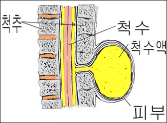
- 척추 후부에 결손이 있고 그 결손 된 구멍을 통해 수막과 척수 신경이 탈출되는 이분 척추를 수막류라고 한다.
- 대부분의 수막류는 정상 피부로 덥혀 있는 것이 보통이다.
- 대개 척수 손상이나 뇌 손상은 없다.
- 때로는 두개골에 생긴 수막류는 뇌수종을 동반 할 수 있고 척추 수막류가 있을 때 배변이나 배뇨 이상이 생길 수 있다.
- 여아에게 척추 수막류가 있으면 직장관 속과 질강 속 사이에 누관이 생길 수 있고 질강이 두개로 갈라져 있을 수 있다.
- 수막류에서 뇌척수액이 새면 뇌막염에 걸리기 쉽다.
- 검진을 하고 적절한 X-선사진 검사, CT 검사, MRI 검사로 진단 할 수 있다.
- 외과적 수술로 치료 한다.
3. 척수 수막류 Myelomeningocele

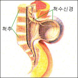
그림 1-101. 척수 수막류
출처: Used with permission from Ross Lab Columbus Ohio,USA와 소아가정간호백과

그림 1-102. 척수 수막류
Copyright ⓒ 2013 John Sangwon Lee, M.D. FAAP
- 척수의 일부가 이분 척추 부분(척추 피열/Spina bifida)을 통해 탈출되어 있는 이분 척추를 척수 수막류라고 한다.
- 태아 발육 과정에서 신경관의 좌우 양 부분이 정상적으로 융합되지 않아 생기는 선천성 신경계 기형의 일종이다.
- 척수, 척수 신경 운동 근육, 수막, 척추, 피부에 이상이 함께 생겨 있을 수 있다. p.00 수두증과 선천성 뇌척수 기형 참조
- 척수 수막류가 있으면 신경 장애, 감각 장애, 근육 마비 등이 척수 수막류 아래 척수신경으로 부터 통제받는 신체 근육에 심각하게 생길 수 있다.
- 신생아 1000명 중 1명에게 나타난다.
- 원인은 확실히 모른다.
- 유전, 임신부의 영양상태, 임신 중 비타민 결핍, 특히 엽산 결핍증, 환경, 약물 등이 원인이 될 수 있다.
척수 수막류의 증상 징후
- 척수 수막류는 이론적으로 어느 척추에도 생길 수 있다.
- 척수 수막류가 척주의 어느 부위에 있느냐에 따라 증상 징후가 다르다.
- 일반적으로 요부 척추와 천골 척추에 가장 많이 생긴다.
- 약 75%는 요부 척추와 천골 척추 부위에 생긴다.
- 드물게는 흉부척추에도 생길 수 있다.
- 흉부 척추에 생기면 흉부척추 이하 운동신경장애, 자율신경 장애, 감각신경 장애가 생겨 심한 장애자가 될 수 있다.
- 요부척추와 천골부척추의 부위에 척수 수막류가 생기면 그 이하 수의근이 마비되고 촉각, 통증 감각이 상실되고 하지에 기형이 생기고 소변과 대변을 가리지 못한다.
- 병력, 증상 징후, 진찰소견을 종합해서 필요한 검사를 해서 진단한다.
- 소아 신경내과 전문의, 소아 신경외과 전문의, 일반 소아청소년과 전문의, 물리치료사, 사회복지사 등이 한 치료 팀이 되어 협동치료한다.
- 임신 초기에 엽산을 충분히 섭취하면 이분척추나 신경관 기형을 예방될 수 있다고 한다.
Spina bifida, Spina bifida occulta, Meningocele and Myelomeningocele
Types of Spina bifida
o The spina bifida occulta
o Meningocele
o Myelomeningocele
• This type of congenital anomaly is caused by vertebral fusion disorder (Dysraphism).
1. Latent bifid vertebrae (closed bifid vertebrae) Spina bifida occulta
• It can occur in any vertebrae, but it most commonly occurs in the fifth lumbar vertebrae of the lumbar region or the first vertebrae of the sacrum.
• There is a very small gap (heat) in the spine due to spinal fusion disorder.
• Among the above three types of diverticulum (Spina bifida), the latent diverticulum is the most common.
• In the presence of latent bipolar vertebrae, there may be more hair, darker skin color, or skin sinuses in the area of the fifth vertebra of the lumbar vertebrae or the first vertebra of the sacral vertebra.
• In most cases, there are no abnormalities in the spinal cord, meninges, nerve roots, or sensation, and there are no symptoms and no health problems.
• Rarely, there may be malformations such as syringomyelia or spina bifida.
• It can cause symptoms.
2. Meningocele
• A bipartite vertebra in which there is a defect in the back of the vertebrae and the meninges and spinal nerves escape through the defect is called meningiomas.
• Most meningiomas are usually covered with normal skin.
• There is usually no spinal cord or brain damage.
• Occasionally, meningiomas of the skull may accompany hydrocephalus, and the presence of spinal meninges may cause defecation or urination abnormalities.
• If a girl has spinal meningitis, a fistula may develop between the rectal canal and the vaginal cavity and the vaginal cavity may split in two.
• Leakage of cerebrospinal fluid from meninges can lead to meningitis.
• Diagnosis can be made through examination and appropriate X-ray examination, CT examination, and MRI examination.
• Treat with surgery.
3. Myelomeningocele

Figure 1-101. spinal meningitis Source: Used with permission from Ross Lab Columbus Ohio, USA and Encyclopedia of Pediatric and Family Nursing

Figure 1-102. spinal meningitis Copyright ⓒ 2013 John Sangwon Lee, M.D. FAAP
A bipartite vertebra in which a portion of the spinal cord prolapses through a bipartite vertebral segment (Spina bifida) is called a spinal meningocele.
• It is a type of congenital anomaly of the nervous system that occurs when the left and right parts of the neural tube do not converge normally during fetal development.
• There may be abnormalities in the spinal cord, spinal nerve motor muscles, meninges, spine, and skin. see Hydrocephalus and Congenital CSF
• With Myelomeningocele, nerve disorders, sensory disturbances, and muscle paralysis can be severe in the muscles of the body controlled by the spinal nerves below the spinal meninges.
• It occurs in 1 in 1000 newborns. The cause is unknown.
• Heredity, nutritional status of the pregnant woman, vitamin deficiency during pregnancy, especially folic acid deficiency, environment, and medications can be the cause.
Symptoms, signs of Myelomeningocele
• Spinal meningiomas can theoretically occur in any vertebra.
• Signs of symptoms vary depending on where in the spinal column the spinal meningeal aneurysm is located.
• It is most common in the lumbar and sacral vertebrae.
• About 75% of these occur in the lumbar and sacral vertebrae.
• Rarely, it can also occur in the thoracic spine.
• If it occurs in the thoracic vertebrae, it can cause sub-thoracic motor neuron disorders, autonomic disorders, and sensory nerve disorders, which can lead to severe disabilities.
• When spinal meninges occur in the lumbar and sacral vertebrae, the lower voluntary muscles are paralyzed, the sense of touch and pain is lost, the lower extremities are deformed, and it is difficult to pass urine and feces.
• Diagnosis is made by taking the necessary tests by synthesizing the medical history, symptom signs, and examination findings.
• Pediatric neurosurgeons, pediatric neurosurgeons, general pediatricians, physical therapists, and social workers work together as one treatment team.
• It is said that a sufficient intake of folic acid in early pregnancy can prevent spina bifida and neural tube malformations.
출처와 참조 문헌 Sources and references
- NelsonTextbook of Pediatrics 22ND Ed
- The Harriet Lane Handbook 22ND Ed
- Growth and development of the children
- Red Book 32nd Ed 2021-2024
- Neonatal Resuscitation, American Academy Pediatrics
- www.drleepediatrics.com 제1권 소아청소년 응급 의료
- www.drleepediatrics.com 제2권 소아청소년 예방
- www.drleepediatrics.com 제3권 소아청소년 성장 발육 육아
- www.drleepediatrics.com 제4권 모유,모유수유, 이유
- www.drleepediatrics.com 제5권 인공영양, 우유, 이유식, 비타민, 미네랄, 단백질, 탄수화물, 지방
- www.drleepediatrics.com 제6권 신생아 성장 발육 육아 질병
- www.drleepediatrics.com제7권 소아청소년 감염병
- www.drleepediatrics.com제8권 소아청소년 호흡기 질환
- www.drleepediatrics.com제9권 소아청소년 소화기 질환
- www.drleepediatrics.com제10권. 소아청소년 신장 비뇨 생식기 질환
- www.drleepediatrics.com제11권. 소아청소년 심장 혈관계 질환
- www.drleepediatrics.com제12권. 소아청소년 신경 정신 질환, 행동 수면 문제
- www.drleepediatrics.com제13권. 소아청소년 혈액, 림프, 종양 질환
- www.drleepediatrics.com제14권. 소아청소년 내분비, 유전, 염색체, 대사, 희귀병
- www.drleepediatrics.com제15권. 소아청소년 알레르기, 자가 면역질환
- www.drleepediatrics.com제16권. 소아청소년 정형외과 질환
- www.drleepediatrics.com제17권. 소아청소년 피부 질환
- www.drleepediatrics.com제18권. 소아청소년 이비인후(귀 코 인두 후두) 질환
- www.drleepediatrics.com제19권. 소아청소년 안과 (눈)질환
- www.drleepediatrics.com 제20권 소아청소년 이 (치아)질환
- www.drleepediatrics.com 제21권 소아청소년 가정 학교 간호
- www.drleepediatrics.com 제22권 아들 딸 이렇게 사랑해 키우세요
- www.drleepediatrics.com 제23권 사춘기 아이들의 성장 발육 질병
- www.drleepediatrics.com 제24권 소아청소년 성교육
- www.drleepediatrics.com 제25권 임신, 분만, 출산, 신생아 돌보기
- Red book 29th-31st edition 2021
- Nelson Text Book of Pediatrics 19th- 21st Edition
- The Johns Hopkins Hospital, The Harriet Lane Handbook, 22nd edition
- 응급환자관리 정담미디어
- Pediatric Nutritional Handbook American Academy of Pediatrics
- 소아가정간호백과–부모도 반의사가 되어야 한다, 이상원 저
- The pregnancy Bible. By Joan stone, MD. Keith Eddleman, MD
- Neonatology Jeffrey J. Pomerance, C. Joan Richardson
- Preparation for Birth. Beverly Savage and Dianna Smith
- 임신에서 신생아 돌보기까지. 이상원
- Breastfeeding. by Ruth Lawrence and Robert Lawrence
- Sources and references on Growth, Development, Cares, and Diseases of Newborn Infants
- Emergency Medical Service for Children, By Ross Lab. May 1989. p.10
- Emergency care, Harvey Grant and Robert Murray
- Emergency Care Transportation of Sick and Injured American Academy of Orthopaedic Surgeons
- Emergency Pediatrics A Guide to Ambulatory Care, Roger M. Barkin, Peter Rosen
- Quick Reference To Pediatric Emergencies, Delmer J. Pascoe, M.D., Moses Grossman, M.D. with 26 contributors
- Neonatal resuscitation Ameican academy of pediatrics
- Pediatric Nutritional Handbook American Academy of Pediatrics
- Pediatric Resuscitation Pediatric Clinics of North America, Stephen M. Schexnayder, M.D.
-
Pediatric Critical Care, Pediatric Clinics of North America, James P. Orlowski, M.D.
-
Preparation for Birth. Beverly Savage and Dianna Smith
-
Infectious disease of children, Saul Krugman, Samuel L Katz, Ann A.
- 제4권 모유, 모유수유, 이유 참조문헌 및 출처
- 제5권 인공영양, 우유, 이유, 비타민, 단백질, 지방 탄수 화물 참조문헌 및 출처
- 제6권 신생아 성장발육 양호 질병 참조문헌 및 출처
-
Neurology in Pediatrics, P. F. Bray, Yearbook Medical Publishers
-
Behavior Disorders in Children, Bakwin and Bakwin
- 소아과학 대한교과서
- 의학 용어사전 대한 의사 협회
Copyright ⓒ 2014 John Sangwon Lee, MD., FAAP
“부모도 반의사가 되어야 한다”-내용은 여러분들의 의사로부터 얻은 정보와 진료를 대신할 수 없습니다.
“The information contained in this publication should not be used as a substitute for the medical care and advice of your doctor. There may be variations in treatment that your doctor may recommend based on individual facts and circumstances.
“Parental education is the best medicine.